Characteristics Of Eukaryotic Cellular Structures ALevel Biology
DNA is the genetic material of the cell. Ribosomes are molecular machines that synthesize proteins. Despite these similarities, prokaryotes and eukaryotes differ in a number of important ways. A prokaryote is a simple, single-celled organism that lacks a nucleus and membrane-bound organelles.

South Pontotoc Biology Plant and Animal Cell Diagrams
Cell Parts ID Game. Test your knowledge by identifying the parts of the cell. Choose cell type (s): Animal Plant Fungus Bacterium. Choose difficulty: Beginner Advanced Expert. Choose to display: Part name Clue. Play.

Anatomy Of A Model Cell mapasgmaes
Structure and Composition of the Cell Membrane. The cell membrane is an extremely pliable structure composed primarily of two layers of phospholipids (a "bilayer"). Cholesterol and various proteins are also embedded within the membrane giving the membrane a variety of functions described below.
Eukaryotic cell structure diagrams Biological Science Picture
Cell diagrams showing a typical animal cell and plant cell. Image created with Biorender.com. Questions Tips & Thanks Want to join the conversation? Sort by: Top Voted lillianjohnson a year ago has there ever been anything that had both animal and plant cells? • 12 comments ( 67 votes) Barrs Julianna a year ago

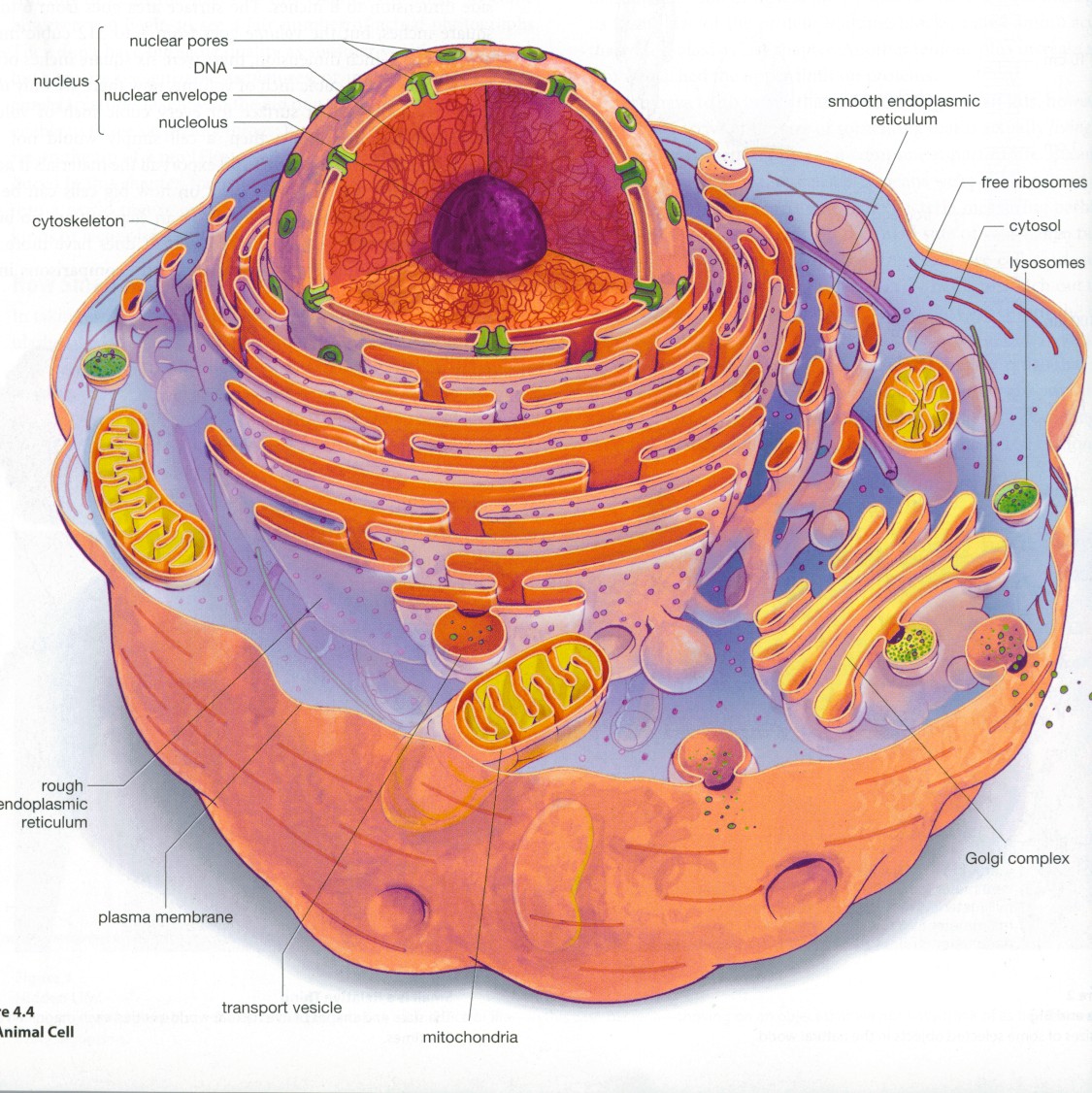
Figure 1.1. Eukaryotic Cell Numerous membranebound organelles are
Unit 1 Intro to biology Unit 2 Chemistry of life Unit 3 Water, acids, and bases Unit 4 Properties of carbon Unit 5 Macromolecules Unit 6 Elements of life Unit 7 Energy and enzymes Unit 8 Structure of a cell Unit 9 More about cells Unit 10 Membranes and transport Unit 11 More about membranes Unit 12 Cellular respiration Unit 13 Photosynthesis

What is a cell? Facts
A diagram of an animal cell is useful for understanding the structure and functioning of an animal. This article includes a well-labeled diagram and a brief description of each component of an animal cell. Animal cells are eukaryotic cells with a membrane-bound nucleus. Since they do not have cell walls and chloroplasts, they are distinct from.

anatomy of the cell
Figure 6.4 Animal cell mitosis is divided into five stages—prophase, prometaphase, metaphase, anaphase, and telophase—visualized here by light microscopy with fluorescence. Mitosis is usually accompanied by cytokinesis, shown here by a transmission electron microscope. (credit "diagrams": modification of work by Mariana Ruiz Villareal; credit "mitosis micrographs": modification of work by.

Biology 101 Cells Owlcation
A cell is the smallest living thing in the human organism, and all living structures in the human body are made of cells. There are hundreds of different types of cells in the human body, which vary in shape (e.g. round, flat, long and thin, short and thick) and size (e.g. small granule cells of the cerebellum in the brain (4 micrometers), up to the huge oocytes (eggs) produced in the female.
Labelled Diagram Of Ribosomes
Label the cell parts you can distinguish. The movement of the cytoplasm is detected by the movement of the chloroplasts which are suspended in cytoplasm. The chloroplasts are carried along in the cytoplasm as if in a current. This movement is known as cyclosis or cytoplasmic streaming. With arrows, indicate the directions of streaming (cyclosis.
Biology Club Our cells 1 ( structure, function, division, disorder
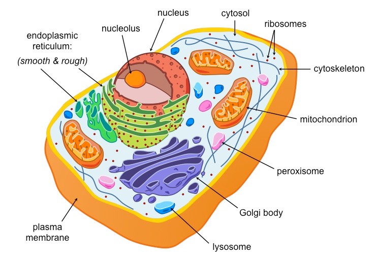
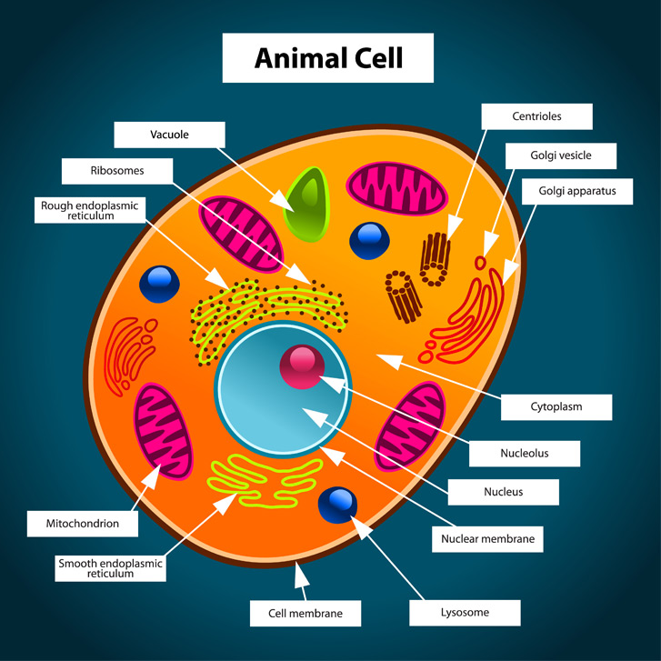
A brief explanation of the different parts of an animal cell along with a well-labelled diagram is mentioned below for reference. Also Read Different between Plant Cell and Animal Cell Well-Labelled Diagram of Animal Cell The Cell Organelles are membrane-bound, present within the cells.

Microtubule Animal Cell
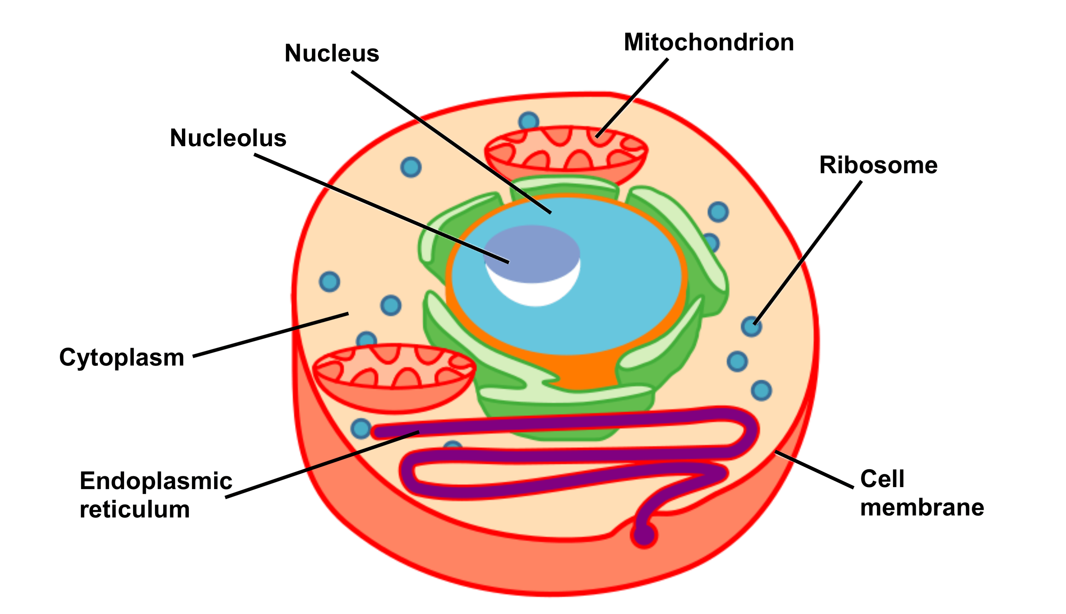
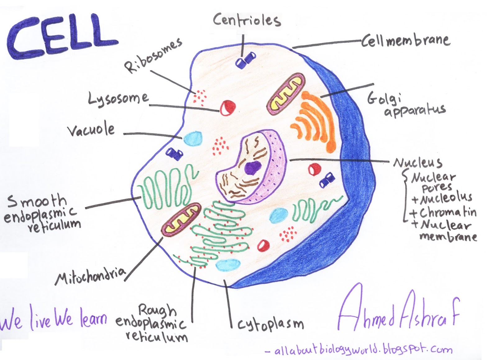
1. Plasma membrane (Cell membrane) 2. Nucleus 3. Cytoplasm 4. Mitochondria 5. Ribosomes 6. Endoplasmic Reticulum (ER) 7. Golgi apparatus (Golgi bodies/Golgi complex) 8. Lysosomes 9. Cytoskeleton Functions of Cytoskeleton 10. Microtubules 11. Centrioles 12. Peroxisomes 13. Cilia and Flagella
Top 158 + Animal cell step by step
What exactly is its job? The plasma membrane not only defines the borders of the cell, but also allows the cell to interact with its environment in a controlled way. Cells must be able to exclude, take in, and excrete various substances, all in specific amounts.

Cells
Animal Cell: Structure, Parts, Functions, Labeled Diagram June 6, 2023 by Faith Mokobi Edited By: Sagar Aryal An animal cell is a eukaryotic cell that lacks a cell wall, and it is enclosed by the plasma membrane. The cell organelles are enclosed by the plasma membrane including the cell nucleus.

The animal cell diagram. Vector illustration on white Etsy in 2021
A membrane-bound nucleus, a central cavity surrounded by membrane that houses the cell's genetic material. A number of membrane-bound organelles, compartments with specialized functions that float in the cytosol.

Plant Cell Diagram Labeled Class 9 Labeled Functions and Diagram
Animal cells are eukaryotic cells, meaning they possess a nucleus and other membrane-bound organelles. Unlike plant cells, animal cells do not have cell walls, allowing for more flexibility in shape and movement. A plasma membrane encloses the cell contents of both plant and animal cells, but it is the outer coating of an animal cell.

Printable Plant Cell Diagram Labeled Printable Diagram Cell diagram
They're one of two major classifications of cells - eukaryotic and prokaryotic. They're also the more complex of the two. Eukaryotic cells include animal cells - including human cells - plant cells, fungal cells and algae. Eukaryotic cells are characterized by a membrane-bound nucleus.